Physical Description
Placida dendritica is a small opisthobranch mollusc. Specimens collected from Heron Island ranged from 5 to 7mm in length, however, individuals over 10mm have been recorded (Thompson, 1976). As a rule, individuals are typically eight times the length of their foot width when fully extended (see Ventral Surface below) (Thompson, 1988). The following description of specimens obtained from Heron Island match descriptions by Alder and Hancock (1845) and Thompson (1976).
Dorsal Surface
Placida dendritica, like all opisthobranch sea slugs, has lost the hard shell that is characteristic of most molluscs. Instead, P. dendritica has a soft, elongate body, with up to 8 transverse rows of typically 3 or 4 cerata (long projections) situated laterally down its dorsal surface (see Figure 1 below). The number and length of cerata is positively correlated with body size (Trowbridge, 1996). The head is relatively unembellished, having only two prominent projections, the rhinophores. The rhinophores are inrolled, and serve a chemosensory function, sensing dissolved chemicals in the seawater. P. dendritica have small, well-developed black eyes, which are located on the head, posterior to the base of the rhinophores. The rhinophores and cerata are soft and smooth like the rest of the body.
Placida dendritica has translucent skin that appears white in colour, and is patterned with a network of green channels (colour varies between individuals, from a dark to light green). These channels are in fact the many branches of the dendritic digestive gland. The green colouration is a result of the retention of chloroplasts from ingested algae in digestive epithelial tissues. All channels branch from two well-defined primary channels running anterior-posteriorly down the animal's dorsal surface (see Figure 1 and Photo (a)). The extensive network of tributaries running off these primary channels spread predominantly into the cerata, but also into the head and rhinophores. This colouration allows these animals to camouflage against the green algal hosts that they feed on. White pigment spots can also be seen all over the body of P. dendritica, however, these are mostly concentrated at the ends of cerata.
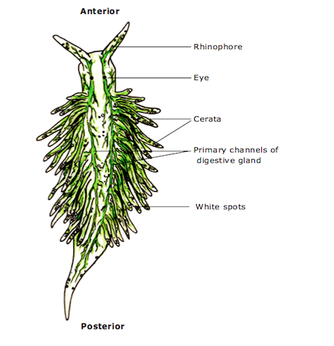
Figure 1:
Dorsal View diagram. Illustration adapted by Alison Carlisle from drawing by Ian F. Smith (2012) and photos taken by A. Carlisle of specimens on Heron Island. Key body characteristics are labelled in this diagram. Comparison of labelled features can be made with those in Photo (a) below.
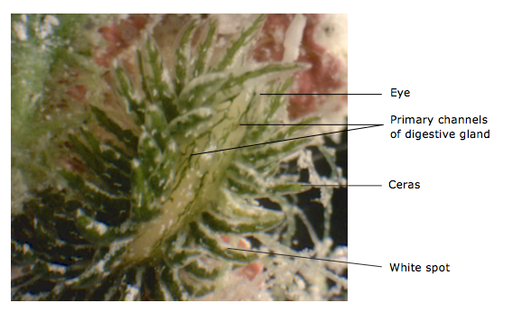 Photo (a):
 Dorsal view of a Placida dendritica specimen from Heron Island, Australia. An eye, ceras (plural: cerata), white pigment spot, and the two primary channels of the digestive system that can be seen through the translucent skin are labelled. Photo taken by Alison Carlisle.
Ventral Surface
The ventral surface of P. dendritica is smooth and translucent-white in colour, interspersed with white pigment spots (see Figure 2). This underside is the animal’s muscular foot, which is used in locomotion. The anterior end (head) is rounded and the posterior end (tail) tapers into a rounded point. The foot is simple and relatively unadorned, with only a small lateral expansion in the middle.

Figure 2: Ventral View diagram. Illustration drawn by Alison Carlisle from photos taken of specimens on Heron Island. Key body characteristics are labelled in this diagram.
|